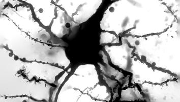
Over a century ago…
Golgi and Cajal revolutionized the field of neuroscience.
For an intriguing summary of this revolution, read Mitch Glickstein’s essay on Golgi and Cajal in Current Biology.

Image credit: I created this serial LM reconstruction of a Golgi-stained mouse pyramidal neuron using a Zeiss Photomicroscope II at 40x oil and Smart Objects in Photoshop.
A new revolution…
Once again neuroscience is undergoing another revolution with the serial electron microscope brain reconstructions.
Petabytes of data have been generated for small volumes (1 cubic mm or less) of human, mouse, songbird, and fly brain volumes. This gargantuan task has been spearheaded by investigators from a variety of entities, including Allen Institute, Baylor, Columbia, Google Research, Harvard, IARPA, Janelia/HHMI, Johns Hopkins, Max Planck Institute, Princeton, Rice, and the University of Cambridge, among others.
Layer 2/3 EM volume: View my analysis of serial electron microscope reconstructed volume of mouse cortex on my Github Layer 2/3 volume repo.
Top image credit: I created this reconstruction using Neuroglancer of pyramidal neuron with cellid 648518346349538440 in the Layer 2/3 volume. See Citation for attribution. Used under Creative Commons Attribution 4.0 International Licence.